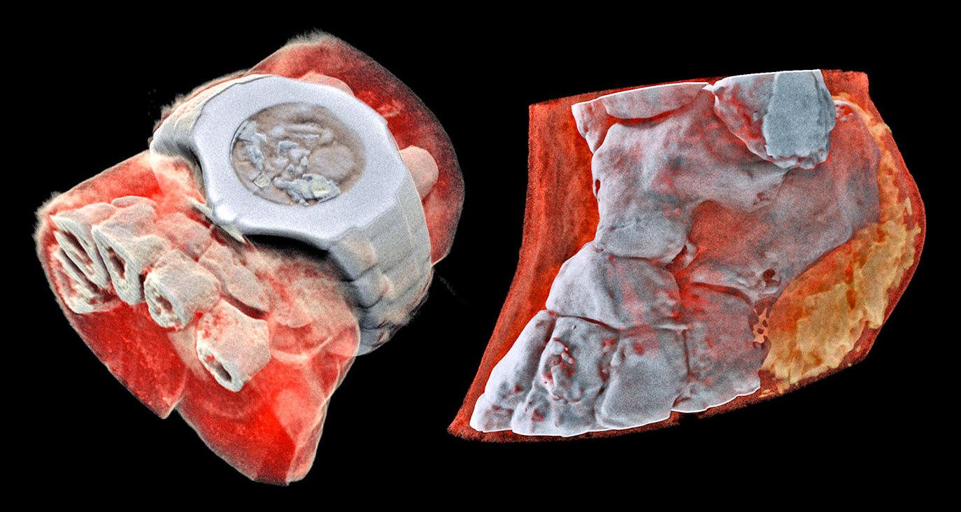
Color x-ray
MARS Bioimaging presented a technique for color and three-dimensional radiography. Instead of black-and-white photos of the insides of the body, which are not always clear to non-specialists, we get a completely new quality thanks to this. Color images not only look fascinating, but also allow doctors to see more than traditional X-rays.
The new type of scanner uses Medipix technology — using computer algorithms and pioneered by scientists at the European Organization for Nuclear Research (CERN) — to track particles at the Large Hadron Collider. Instead of registering X-rays as they pass through tissues and how they are absorbed, the scanner determines the exact energy level of the radiation as it hits various parts of the body. It then converts the results into different colors to match bones, muscles, and other tissues.
The MARS scanner is already being used in many studies, including cancer and stroke studies. Now the developers want to test their equipment in the treatment of orthopedic and rheumatological patients in New Zealand. However, even if all goes well, it may be many years before the camera is properly certified and approved for normal medical use.