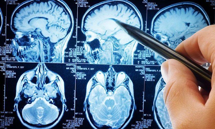
medical imaging
In 1896, Wilhelm Roentgen discovered X-rays, and in 1900, the first chest X-ray. Then comes the X-ray tube. And what it looks like today. You will find out in the article below.
1806 Philippe Bozzini develops the endoscope in Mainz, publishing on the occasion "Der Lichtleiter" - a textbook on the study of the recesses of the human body. The first to use this device in a successful operation was the Frenchman Antonin Jean Desormeaux. Before the invention of electricity, external light sources were used to examine the bladder, uterus, and colon, as well as the nasal cavities.
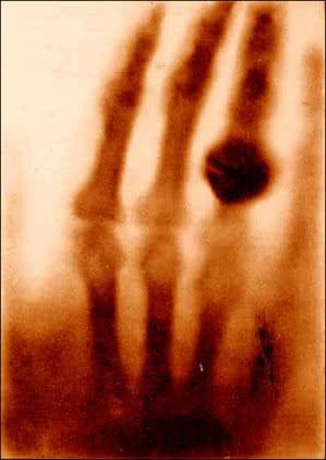
1. The first X-ray - the hand of Roentgen's wife
1896 Wilhelm Roentgen discovers X-rays and their ability to penetrate solids. The first specialists to whom he showed his "roentgenograms" were not doctors, but Roentgen's colleagues - physicists (1). The clinical potential of this invention was recognized a few weeks later, when an X-ray of a shard of glass in the finger of a four-year-old child was published in a medical journal. Over the next few years, the commercialization and mass production of X-ray tubes spread the new technology around the world.
1900 First chest x-ray. The widespread use of chest x-rays made it possible to detect tuberculosis at an early stage, which at that time was one of the most common causes of death.
1906-1912 The first attempts to use contrast agents for better examination of organs and vessels.
1913 A real X-ray tube, called a hot cathode vacuum tube, is emerging, which uses an efficient controlled electron source due to the phenomenon of thermal emission. He opened a new era in medical and industrial radiological practice. Its creator was the American inventor William D. Coolidge (2), popularly known as the "father of the X-ray tube." Together with the moveable grid created by Chicago radiologist Hollis Potter, the Coolidge lamp made radiography an invaluable tool for physicians during World War I.
1916 Not all radiographs were easy to read - sometimes tissues or objects obscured what was being examined. Therefore, the French dermatologist André Bocage developed a method of emitting X-rays from different angles, which eliminated such difficulties. His .
1919 Pneumoencephalography appears, which is an invasive diagnostic procedure of the central nervous system. It consisted in replacing part of the cerebrospinal fluid with air, oxygen or helium, introduced through a puncture into the spinal canal, and performing an x-ray of the head. The gases were well contrasted with the ventricular system of the brain, which made it possible to obtain an image of the ventricles. The method was widely used in the middle of the 80th century, but was almost completely abandoned in the XNUMXs, since the examination was extremely painful for the patient and was associated with a serious risk of complications.
30s and 40s In physical medicine and rehabilitation, the energy of ultrasonic waves is beginning to be widely used. Russian Sergey Sokolov is experimenting with the use of ultrasound to find metal defects. In 1939, he uses a frequency of 3 GHz, which, however, does not provide satisfactory image resolution. In 1940, Heinrich Gohr and Thomas Wedekind of the Medical University of Cologne, Germany, presented in their article "Der Ultraschall in der Medizin" the possibility of ultrasound diagnostics based on echo-reflex techniques similar to those used in the detection of metal defects. .
The authors hypothesized that this method would allow the detection of tumors, exudates, or abscesses. However, they could not publish convincing results of their experiments. Also known are the ultrasonic medical experiments of the Austrian Karl T. Dussik, a neurologist from the University of Vienna in Austria, started in the late 30s.
1937 The Polish mathematician Stefan Kaczmarz formulates in his work "Technique of Algebraic Reconstruction" the theoretical foundations of the method of algebraic reconstruction, which was then applied in computed tomography and digital signal processing.
40-s years. The introduction of a tomographic image using an x-ray tube rotated around the patient's body or individual organs. This made it possible to see the details of the anatomy and pathological changes in the sections.
1946 American physicists Edward Purcell and Felix Bloch independently invented nuclear magnetic resonance NMR (3). They were awarded the Nobel Prize in Physics for "the development of new methods of precise measurement and related discoveries in the field of nuclear magnetism."
3. Set of NMR equipment
1950 rises rectilinear scanner, compiled by Benedict Cassin. The device in this version was used until the early 70s with various radioactive isotope-based pharmaceuticals to image organs throughout the body.
1953 Gordon Brownell of the Massachusetts Institute of Technology creates a device that is the forerunner of the modern PET camera. With her help, he, along with neurosurgeon William H. Sweet, manages to diagnose brain tumors.
1955 Dynamic x-ray image intensifiers are being developed that make it possible to obtain x-ray images of moving images of tissues and organs. These x-rays have provided new information about bodily functions such as the beating heart and the circulatory system.
1955-1958 Scottish doctor Ian Donald begins to widely use ultrasound tests for medical diagnosis. He is a gynecologist. His article "Investigation of Abdominal Masses with Pulsed Ultrasound", published on June 7, 1958 in the medical journal The Lancet, defined the use of ultrasound technology and laid the foundation for prenatal diagnosis (4).
1957 The first fiber optic endoscope is developed - gastroenterologist Basili Hirshowitz and his colleagues from the University of Michigan patent a fiber optic, semi-flexible gastroscope.
1958 Hal Oscar Anger presents at the annual meeting of the American Society for Nuclear Medicine a scintillation chamber that allows for dynamic imaging of human organs. The device enters the market after a decade.
1963 Freshly minted Dr. David Kuhl, together with his friend, engineer Roy Edwards, present to the world the first joint work, the result of several years of preparation: the world's first apparatus for the so-called. emission tomographywhich they call the Mark II. In subsequent years, more accurate theories and mathematical models were developed, numerous studies were carried out, and more and more advanced machines were built. Finally, in 1976, John Keyes creates the first SPECT machine - single photon emission tomography - based on the experience of Cool and Edwards.
1967-1971 Using the algebraic method of Stefan Kaczmarz, English electrical engineer Godfrey Hounsfield creates the theoretical foundations of computed tomography. In the following years, he constructs the first working EMI CT scanner (5), on which, in 1971, the first examination of a person is carried out at the Atkinson Morley Hospital in Wimbledon. The device was put into production in 1973. In 1979, Hounsfield, along with the American physicist Allan M. Cormack, was awarded the Nobel Prize for their contribution to the development of computed tomography.
5. EMI Scanner
1973 The American chemist Paul Lauterbur (6) discovered that by introducing gradients of a magnetic field passing through a given substance, one can analyze and find out the composition of this substance. The scientist uses this technique to create an image that distinguishes between normal and heavy water. Based on his work, English physicist Peter Mansfield builds his own theory and shows how to make a quick and accurate image of the internal structure.
The result of the work of both scientists was a non-invasive medical examination, known as magnetic resonance imaging or MRI. In 1977, the MRI machine, developed by American physicians Raymond Damadian, Larry Minkoff and Michael Goldsmith, was first used to study a person. Lauterbur and Mansfield were jointly awarded the 2003 Nobel Prize in Physiology or Medicine.
1974 American Michael Phelps is developing a Positron Emission Tomography (PET) camera. The first commercial PET scanner was created thanks to the work of Phelps and Michel Ter-Poghosyan, who led the development of the system at EG&G ORTEC. The scanner was installed at UCLA in 1974. Because cancer cells metabolize glucose ten times faster than normal cells, malignant tumors appear as bright spots on a PET scan (7).
1976 Surgeon Andreas Grünzig presents coronary angioplasty at the University Hospital Zurich, Switzerland. This method uses fluoroscopy to treat blood vessel stenosis.
1978 rises digital radiography. For the first time, an image from an X-ray system is converted into a digital file, which can then be processed for a clearer diagnosis and stored digitally for future research and analysis.
80-s years. Douglas Boyd introduces the method of electron beam tomography. EBT scanners used a magnetically controlled beam of electrons to create a ring of X-rays.
1984 The first 3D imaging using digital computers and CT or MRI data appears, resulting in XNUMXD images of bones and organs.
1989 Spiral computed tomography (spiral CT) comes into use. This is a test that combines a continuous rotational movement of the lamp-detector system and movement of the table over the test surface (8). An important advantage of spiral tomography is the reduction of the examination time (it allows you to obtain an image of several dozen layers in one scan lasting several seconds), the collection of readings from the entire volume, including the layers of the organ, which were between scans with traditional CT, as well as the optimal transformation of the scan thanks to new software . The pioneer of the new method was Siemens Director of Research and Development Dr. Willy A. Kalender. Other manufacturers soon followed in the footsteps of Siemens.
8. Scheme of spiral computed tomography
1993 Develop an echoplanar imaging (EPI) technique that will allow MRI systems to detect acute stroke at an early stage. EPI also provides functional imaging of, for example, brain activity, allowing clinicians to study the function of different parts of the brain.
1998 The so-called multimodal PET examinations together with computed tomography. This was done by Dr. David W. Townsend of the University of Pittsburgh, along with Ron Nutt, a PET systems specialist. This has opened up great opportunities for metabolic and anatomical imaging of cancer patients. The first prototype PET/CT scanner, designed and built by CTI PET Systems in Knoxville, Tennessee, went live in 1998.
2018 MARS Bioimaging introduces the color i technique XNUMXD medical imaging (9), which, instead of black and white photographs of the inside of the body, offers a completely new quality in medicine - color images.
The new type of scanner uses Medipix technology, first developed for scientists at the European Organization for Nuclear Research (CERN) to track particles at the Large Hadron Collider using computer algorithms. Instead of recording X-rays as they pass through tissues and how they are absorbed, the scanner determines the exact energy level of X-rays as they hit different parts of the body. It then converts the results into different colors to match bones, muscles, and other tissues.
9. Colored section of the wrist, made using MARS Bioimaging technology.
Classification of medical imaging
1. X-ray (X-ray) this is an x-ray of the body with the projection of x-rays onto a film or detector. Soft tissues are visualized after contrast injection. The method, which is mainly used in the diagnosis of the skeletal system, is characterized by low accuracy and low contrast. In addition, radiation has a negative effect - 99% of the dose is absorbed by the test organism.
2. tomography (Greek - cross section) - the collective name of diagnostic methods, which consist in obtaining an image of a cross section of a body or part of it. Tomographic methods are divided into several groups:
- UZI (UZI) is a non-invasive method that uses the wave phenomena of sound at the boundaries of various media. It uses ultrasonic (2-5 MHz) and piezoelectric transducers. The image moves in real time;
- computed tomography (CT) uses computer-controlled x-rays to create images of the body. The use of x-rays brings CT closer to x-rays, but x-rays and computed tomography provide different information. It is true that an experienced radiologist can also infer the three-dimensional location of, for example, a tumor from an X-ray image, but X-rays, unlike CT scans, are inherently two-dimensional;
- magnetic resonance imaging (MRI) - this type of tomography uses radio waves to examine patients placed in a strong magnetic field. The resulting image is based on radio waves emitted by the examined tissues, which generate more or less intense signals depending on the chemical environment. The body image of the patient can be saved as computer data. MRI, like CT, produces XNUMXD and XNUMXD images, but is sometimes a much more sensitive method, especially for distinguishing soft tissues;
- positron emission tomography (PET) - registration of computer images of changes in sugar metabolism occurring in tissues. The patient is injected with a substance that is a combination of sugar and isotopically labeled sugar. The latter makes it possible to locate the cancer, since cancer cells take up sugar molecules more efficiently than other tissues in the body. After ingestion of radioactively labeled sugar, the patient lies down for approx.
- 60 minutes while the marked sugar circulates in his body. If there is a tumor in the body, sugar must be efficiently accumulated in it. Then the patient, laid on the table, is gradually introduced into the PET scanner - 6-7 times within 45-60 minutes. The PET scanner is used to determine the distribution of sugar in body tissues. Thanks to the analysis of CT and PET, a possible neoplasm can be better described. The computer-processed image is analyzed by a radiologist. PET can detect abnormalities even when other methods indicate the normal nature of the tissue. It also makes it possible to diagnose cancer relapses and determine the effectiveness of treatment - as the tumor shrinks, its cells metabolize less and less sugar;
- Single photon emission tomography (SPECT) – tomographic technique in the field of nuclear medicine. With the help of gamma radiation, it allows you to create a spatial image of the biological activity of any part of the patient's body. This method allows you to visualize the blood flow and metabolism in a given area. It uses radiopharmaceuticals. They are chemical compounds consisting of two elements - a tracer, which is a radioactive isotope, and a carrier that can be deposited in tissues and organs and overcome the blood-brain barrier. Carriers often have the property of selectively binding to tumor cell antibodies. They settle in quantities proportional to metabolism;
- optical coherence tomography (OCT) - a new method similar to ultrasound, but the patient is probed with a beam of light (interferometer). Used for eye examinations in dermatology and dentistry. Backscattered light indicates the position of places along the path of the light beam where the refractive index changes.
3. Scintigraphy - we get here an image of organs, and above all their activity, using small doses of radioactive isotopes (radiopharmaceuticals). This technique is based on the behavior of certain pharmaceuticals in the body. They act as a vehicle for the isotope used. The labeled drug accumulates in the organ under study. The radioisotope emits ionizing radiation (most often gamma radiation), penetrating outside the body, where the so-called gamma camera is recorded.